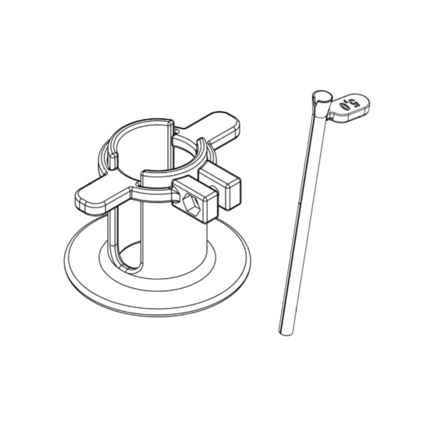
Dilation System
Dilation system that enables the dilation of narrow channels that arise after the standard insertion of thin drains into a cavity, hollow organ, or pathological area.

Dilation system that enables the dilation of narrow channels that arise after the standard insertion of thin drains into a cavity, hollow organ, or pathological area.
Background
Currently, common solid or flexible dilators or braided stents are used to secure and dilate entrances into body cavities (pleural and abdominal), necrotic and infected cavities in the retroperitoneum or into hollow organs through the body surface. The disadvantage of such procedures is the possibility of damaging surrounding tissues and the risk of deformation of the created channel due to the thick subcutaneous layer and muscle during the insertion of the dilator. With braided stents supplemented with dilation balloons, it is very problematic to achieve an uniformly dilated channel and to remove the braided stent due to possible tissue ingrowth and adhesion of the stent to the surroundings. The disadvantage of braided stents is also their high price.
Descricption of the Invention
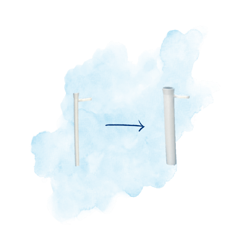
The aim of the invention is to dilate narrow channels that arise after the standard insertion of thin drains into a cavity, hollow organ, or pathological area. The insertion is often done under ultrasound or X-ray control. The newly developed dilatation system is designed to allow the expansion of the channel through which the thin drain is inserted. The uniqueness of the new system lies in the designed dilator set that allows a larger diameter dilator to be inserted into a smaller diameter dilator. This procedure is repeated until a desired width of the channel is achieved. Thus, the channel gradually and gently expands and eventually allows the insertion of thicker drain. The new method of dilation reduces damage to surrounding tissues that might be caused by dilation. The depth of the inserted dilator is determined by a fixation device (fixation ring), which can be used to safely set the depth of the dilated channel.
Advantages and Potential Application
The invention is primarily intended for the insertion of thicker drains in patients with infectious and necrotic deposits in the abdominal cavity, retroperitoneum, or chest cavity. However, it is also suitable for dilating entrances to hollow organs (trachea, stomach, uterus, urinary system). It can also be used during laparoscopic procedures.
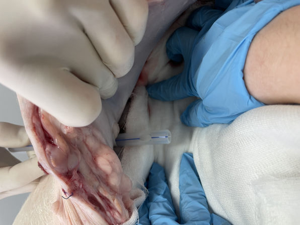
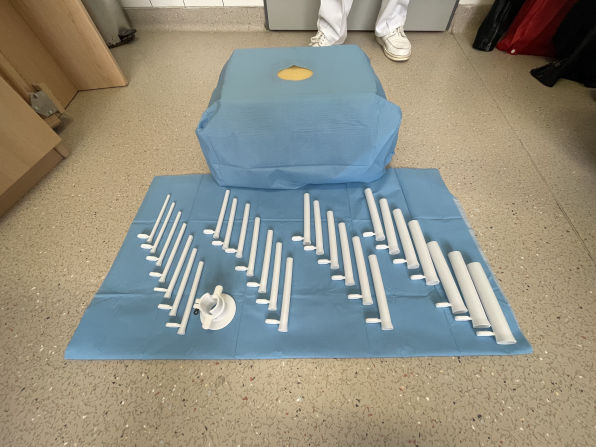
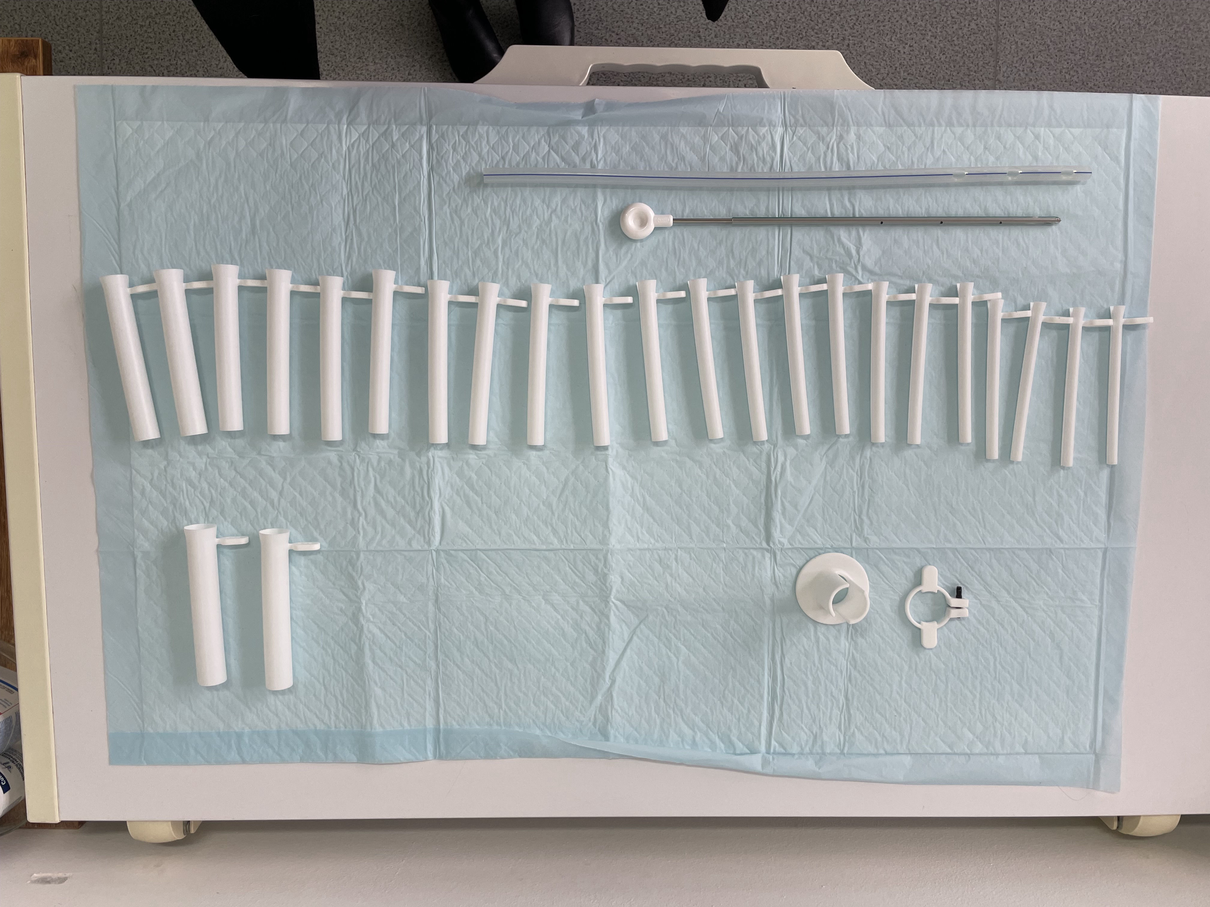
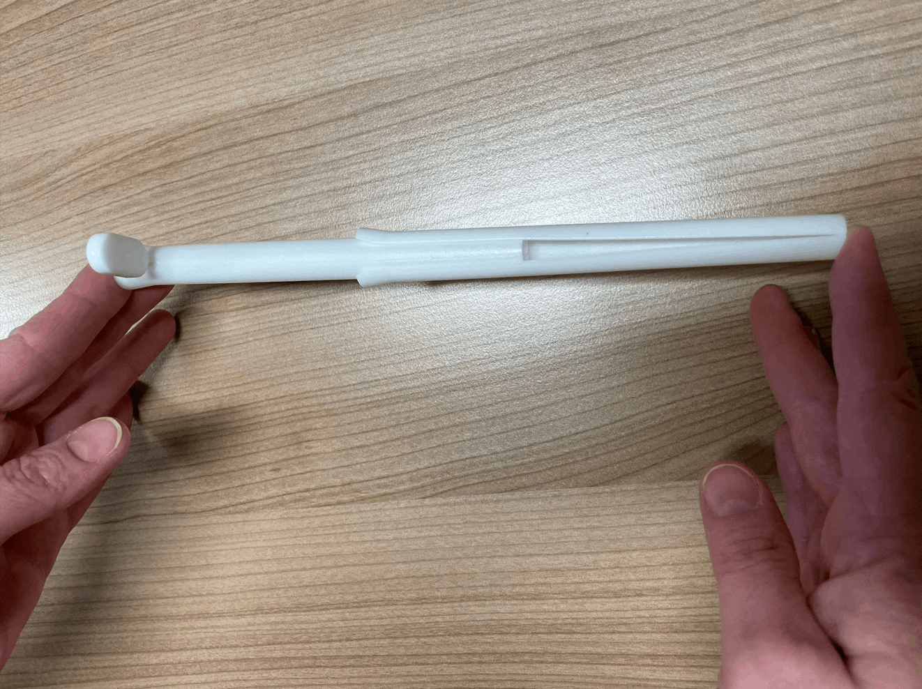
Technology is owned by University Hospital Hradec Králové. Czech patent application filed (priority date 7. 12. 2022). 3D printed set of dilators were tested in vivo in pig model, where all the expected benefits were confirmed by the clinician. The development was supported by the Technology Agency of the Czech Republic within the programme for applied research, experimental development and Innovation GAMA 2 – Project TP01010034 – Centre for Transfer of Biomedical Technologies – PoC2. Now we are searching for commercial partner that would bring our device to the market.

The 3rd Department of Internal Medicine - Metabolic Care and Gerontology
Principal investigator & coinvestigator of multiple research projects and clinical studies
Member of the European Society for Clinical Nutrition and Metabolism, the Czech Society for Clinical Nutrition and Intensive Metabolic Care
Owner of multiple patents & licensed technology
For more detailed information, do not hesitate to contact us directly:
Lucie Bartošová, Ph.D., lucie.bartosova@fnhk.cz or +420 495 832 925.
Stay in touch with us on social media where you can follow recent progress in developing technologies.

Free AI Website Creator